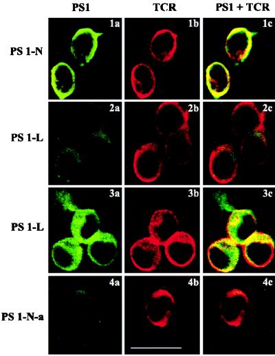
Figure 4.
Immunolocalization of PS1 in Jurkat cells. PS1 (green) and TCR (red) were localized in Jurkat cells by confocal immunofluorescent microscopy. (1a–1c, 2a–2c, and 4a–4c) Nonpermeabilized cells. (3a–3c) Cells permeabilized by 0.5% Triton X-100. Cells were coincubated with mAb to TCR and PS-N (1a–1c), PS-L (2a–2c and 3a–3c), and preabsorbed antibody PS1-N-a (4a–4c). (Right) Colocalization of PS1 with TCR is shown (1c, 2c, 3c, and 4c). (Bar = 10 μm.)

