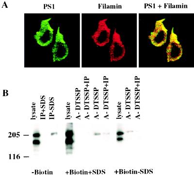
Figure 6.
Interaction of cell-surface PS1 with filamin in Jurkat cells. (A) Colocalization of PS1 and filamin in Jurkat cells. Jurkat cells were plated on chambered coverslips covered with saturated solution of rat tail collagen, type 1. Cells were fixed and permeabilized with 0.5% Triton X-100. PS1 (green) and filamin (red) were visualized in Jurkat cells by multiple optical sectioning (0.2 μ) by using confocal immunofluorescent microscopy. (×1,600.) Antibodies used were anti-N-terminal rabbit antibody (PS1-N) and antifilamin mAb PM 6/317. (B) Binding of cell-surface PS1 to filamin in Jurkat cells. Jurkat cells were treated with biotin (+Biotin) or were not treated (−Biotin). Cells were permeabilized by lysolecithin and treated with the reversible cross-linker (+DTSSP), whereas control cells were not treated with cross-linker (−DTSSP). The cells subsequently were lysed with or without 0.4% SDS (+SDS or −SDS), cell lysates were subjected to chromatography on immobilized monomeric avidin (A), and bound proteins were eluted with 2 mM d-biotin and immunoprecipitated with anti-PS1 N-terminal antibody 231f (IP). Immunoprecipitates were boiled for 10 min in Laemmli sample buffer containing 100 mM 2-mercaptoethanol, separated on a 6% SDS/PAGE gel, and Western blotted with antifilamin mAb PM 6/317. The molecular mass markers are shown on the left.

