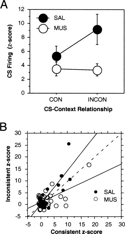
Figure 5.
(A) Mean (±SEM) CS-evoked firing (z-scores) in lateral amygdala neurons 50–100 msec after CS onset after saline (SAL) or muscimol (MUS) infusion into the dorsal hippocampus during the consistent (CON) and inconsistent (INCON) retention tests. (B) Scatterplot of normalized spike firing (50–100 msec after CS onset) for each neuron recorded under SAL (solid circles) or MUS (open circles) in the consistent and inconsistent test sessions. Regression lines indicate the deviation from a slope of 1 (dashed line) under the different drug conditions.

