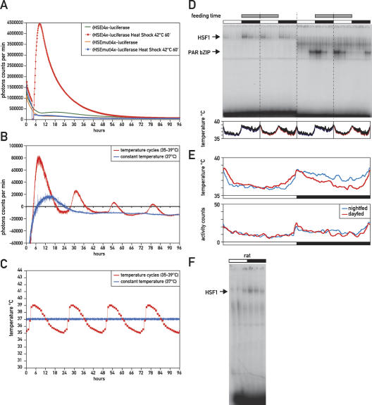
Figure 5.
Body temperature and feeding affect the phase of circadian HSF1 activity. (A) Real-time bioluminescence analysis of the HSE-luciferase reporter plasmids (HSE)4x-Luciferase and (HSEmut)4x-Luciferase in NIH3T3 mouse fibroblasts after a 60-min heat shock at 42°C or at constant temperature. (B) Real-time bioluminescence analysis of the HSE-luciferase reporter plasmid (HSE)4x-Luciferase in NIH3T3 cells that were entrained to temperature cycles (35°C–39°C) or kept at constant temperature (37°C). Luciferase expression of the non-temperature-responsive control plasmid (HSEmut)4x-Luciferase was subtracted, resulting in partially negative values for luciferase expression. (C) Real-time temperature monitoring of the tissue culture medium in the temperature entrainment experiment shown in B. (D) DNA-binding activity of HSF1 and PAR bZIP proteins in mice that were fed exclusively during the night or during the day. Feeding time is indicated by gray bars, while white and black bars indicate light and dark phases, respectively. Sampled real-time temperature measurements in living animals are shown below the autoradiograph. Colored lines indicate sampled body temperature of four night-fed (blue) or day-fed (red) animals during the last 7 d before they were sacrificed. Standard deviations are shown by black error bars on top of the colored lines. (E) The top panel shows an overlay of the sampled body temperature of night-fed (blue) and day-fed (red) animals. The bottom panel shows sampled activity counts for the same animals. (F) Circadian EMSA of HSF1 in rat.

