Figure 1.
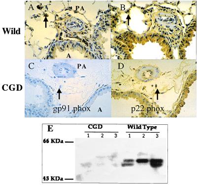
gp91 phox and p22 phox are present in the lung. gp91 phox (A) and p22 phox (B) are present in wild-type mice (immunohistochemistry in brown, counterstain blue). Controls lacking primary antibody are completely negative (not shown). As expected, the gp91 phox (C), but not the p22 phox (D), is absent in the CGD mouse. Lack of gp91 phox in C reveals the hematoxylin counterstain. Qualitative grading of the intensity of expression of gp91 phox by blinded observers was: alveolar macrophages (arrows), 4+; alveolar epithelium airways, 3+; pulmonary veins, 3+; large PAs, 2+; and small PAs, 1+. (E) Immunoblots show a typical band for gp91 phox at ≈60 kDa. The gp91 phox refers to the molecular weight in human tissue, where the subunit is more heavily glycosylated.

