
Fig 9. Raf and ERK scaffolding properties of β-arrestin2, β-arrestin20K and β-arrestin2-Ub.
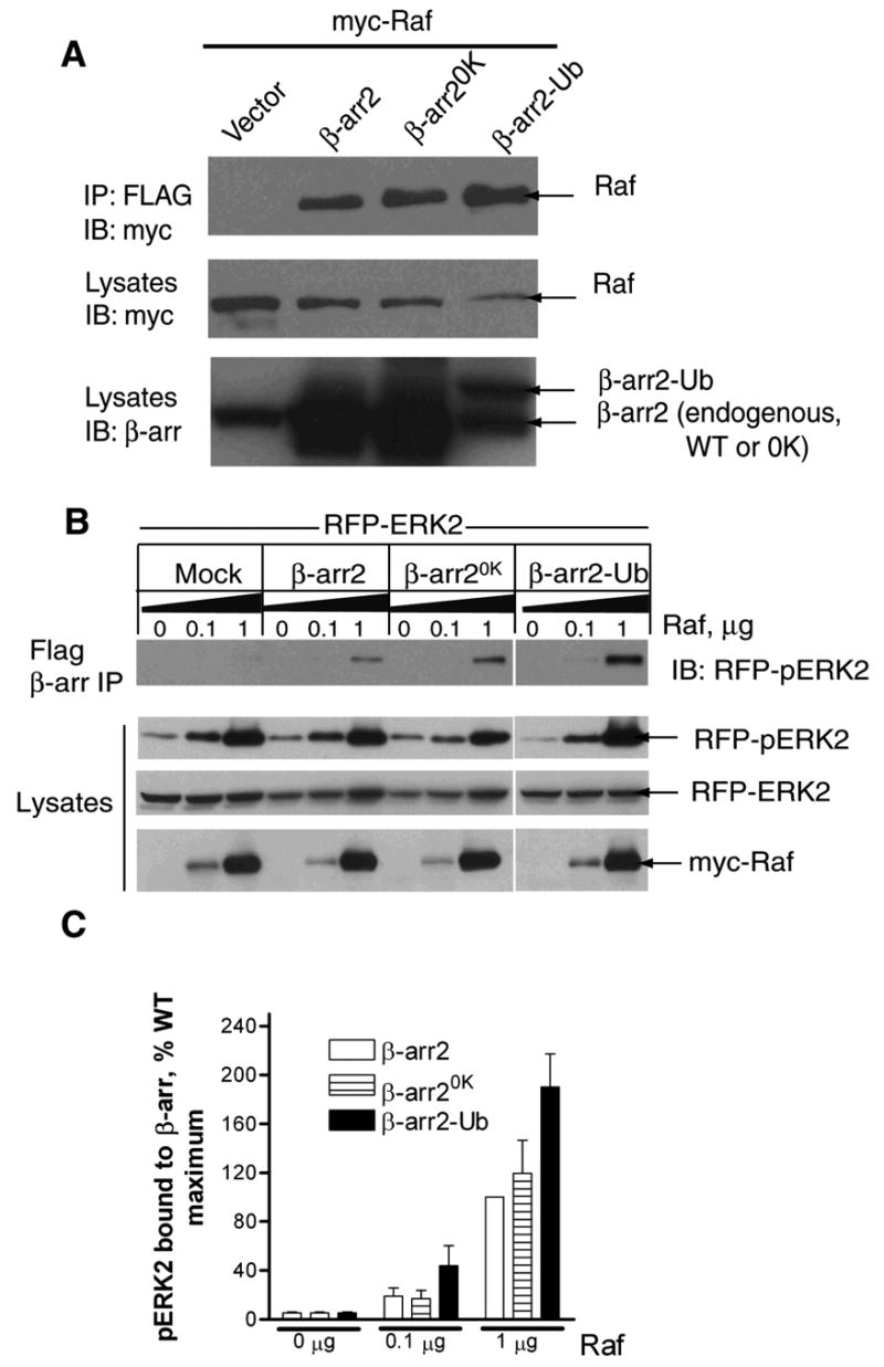
A. COS-7 cells were transfected with myc-cRaf1 along with either vector or the indicated β-arrestin2 plasmid. Anti-FLAG immunoprecipitates were probed with a monoclonal myc antibody (9E10) (top panel). The lysates were probed for the levels of transfected cRaf1 (middle panel) and β-arrestins (bottom panel) with respective antibodies. Blots are representative of four similar experiments. B. Cells were transiently transfected with RFP-ERK2 and increasing amounts of myc-cRaf1 with vector or indicated FLAG-β-arrestins. The amounts of phospho-RFP-ERK2 present in the FLAG immunoprecipitates (top panel) and lysates were determined by immunoblotting. The levels of unphosphorylated RFP-ERK2 as well as myc-cRaf1 in the same lysates are also shown. C. Bar graph depicts the amount of phospho-RFP-ERK2 in the FLAG immunoprecipitates. Data are presented in arbitrary units where the amount of maximum phospho-ERK2 present in FLAG-β-arrestin2 immunoprecipitates is defined as 100%. Data shown represent the mean ± SEM from three independent experiments.