
Figure 8.
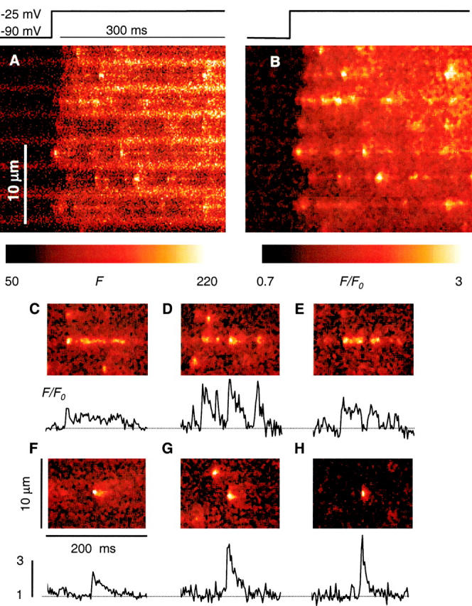
Local effects of high Mg2+. (A and B) Line scan image of fluorescence in a fiber exposed for 115 min to internal solution with 7 mM [Mg2+]. Release was elicited by depolarization to −25 mV, as indicated. (A) Raw fluorescence. (B) F/F 0, after nine-point median filtering. To improve contrast, color scale does not start at 0. Identifier, 0107b. (C–G) Selected events from images in high Mg2+, normalized and median filtered as in B. (H) An event in reference, with characteristics of an average spark. The graphs under each panel are averages over five pixels at the center of the events, all with the same vertical scale. The color table was adjusted in each panel for best visualization of the increase in fluorescence that follows the sparks. Identifiers for C–H: 1106a, 1022a, 0623a, 0107b, 1022a, 0315a.