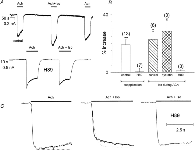
Figure 1.
Effect of β-adrenergic stimulation on IKACh of rat atrial cells. (A) Original whole cell current recordings at a membrane holding potential of −80 mV. IKACh was induced by superfusion with ACh (10 μmol/liter) or ACh plus Iso (1 μmol/liter) in the absence (top) and in the presence (bottom) of 50 μmol/liter H89. (B) Statistics of the effect of Iso on IKACh amplitude, shown as percent increase compared with IKACh induced by ACh alone. Calculated average values ± SEM are shown; the number of individual cells is given in parenthesis. (* and **) Mean value deviates significantly (P < 0.05 and 0.01) from the H89 treated group. (Left to right) Effect of Iso coapplied with ACh (control, H89 treatment); additional IKACh induced by Iso application during ACh (control, IKACh recorded with nystatin perforated patch, H89 treatment). (C) Effect of Iso coapplication on kinetics of I KACh activation. (Left to right) Regular IKACh induced by ACh superfusion; IKACh induced by ACh coapplied with Iso; same as previous, but in the presence of 50 μmol/liter H89. (Dotted line) Monoexponential fit through the rising phase of I KACh. (Broken line) Biexponential fit, comprising a fast and an additional slow time constant.

