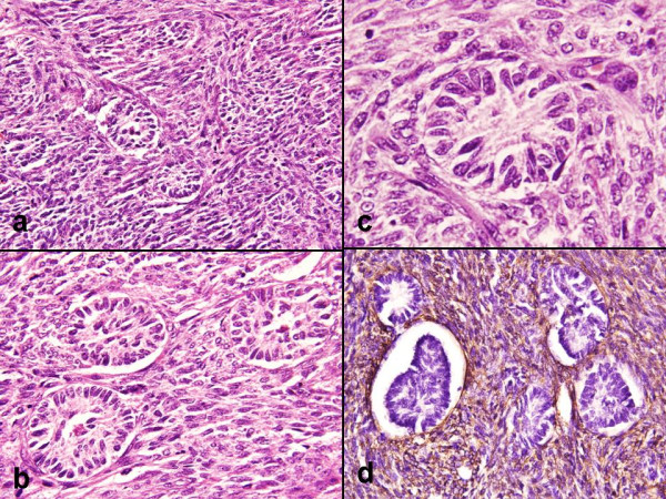
Figure 2.

a). Cellular tumor composed of spindled cells in sheets, fascicles and whorls (HE × 200). b). Small aggregates of undifferentiated sex cord type cells forming poorly defined tubular structure (HE × 300). c). Small aggregates were sharply demarcated from the surrounding stroma (HE × 400). d). IHC, SMA intense cytoplasmic staining in the spindled cells (Avidin biotin staining × 400).
