
Figure 2.
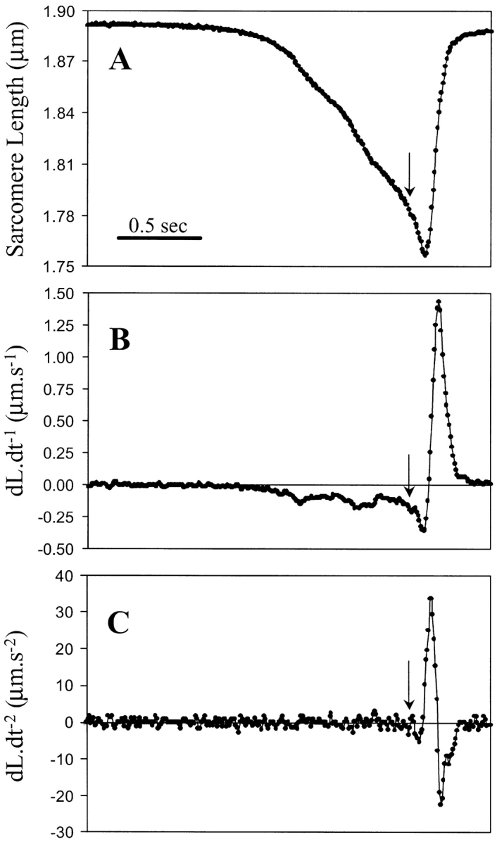
Changes in cell length, and the corresponding first and second derivative of the changes in cell length for a rigor induced contraction and relaxation. (A) At a given time, the perfusate is switched to ATP-free rigor solution, and while the cell is shortening the perfusate is switched back to ATP containing solution (arrow). After ∼150 ms the myocyte starts to relengthen, indicating completion of the perfusate switch and detachment of cross-bridges. (B) First derivative (dL·dt−1) of the tracing shown in A, indicating velocity of shortening and relengthening. (C) Second derivative (dL·dt−2) of the tracing shown in A, indicating acceleration and deceleration of the sarcomere. All tracings were acquired at a sampling rate of 120 Hz.