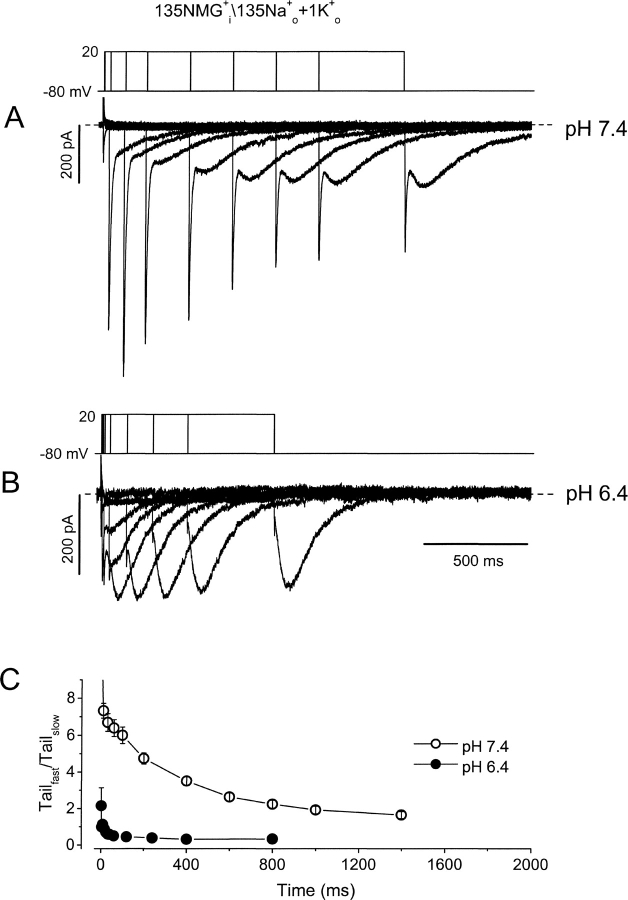
Figure 3.
Extracellular acidification promotes inactivation. (A and B) Superimposed tail currents after depolarizations of varying duration at pH 7.4 and pH 6.4. Pipette contained NMG+ as the major intracellular cation, and the bath solution contained 135 Na+ o + 1 mM K+ o. On the first pulse the membrane was depolarized from −80 to 20 mV for 2 ms at pH 7.4, and 1 ms at pH 6.4. For subsequent pulses the protocol was repeated every 10 s for increasing durations, 10, 30, 60, 100, 200, 400, 600, 800, 1,000, and 1,400 ms at pH 7.4; and 4, 8, 16, 24, 32, 40, 60, 120, 240, 400, and 800 ms at pH 6.4. Tail currents are composed of a fast component, reflecting deactivation from the open state, and a slow component reflecting Na+ current through inactivated channels. (C) The ratio of peak transient inward current amplitude versus the peak slow tail current is plotted against the duration of depolarization at 20 mV at pH 7.4 (○) and pH 6.4 (•).

