Figure 6.
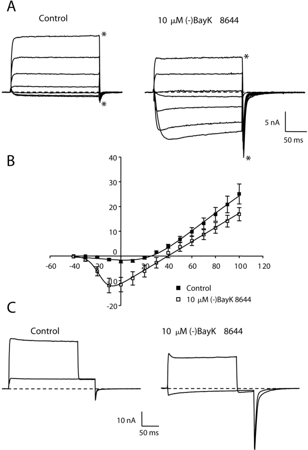
An Mg2+-free pipette solution abolishes voltage-dependent potentiation of inward current. (A) Representative whole-cell currents measured with a Mg2+-free pipette solution (see materials and methods) from a dysgenic myotube expressing E2A/E4A-α1C before (left) and after (right) application of 10 μM (−)BayK 8644. The illustrated currents were elicited by 200-ms depolarizations to potentials ranging from −20 to 100 mV in increments of 20 mV, followed by repolarization to −50 mV. (B) Average peak current versus voltage relationship for E2A/E4A-α1C in the absence (closed symbols, n = 12) or presence (open symbols, n = 9) of 10 μM (−)BayK 8644 with a Mg2+-free pipette solution. The smooth curves represent best fits of the Boltzmann expression (see materials and methods) yielding the values (control; BayK), Gmax = 330 nS/nF; 287 nS/nF, Vrev = 25.7 mV; 39.8 mV, V1/2 = 23.6 mV; −17.4 mV, kG = 16.6 mV; 4.7 mV. (C) Absence of depolarization-induced potentiation of E2A/E4A-α1C currents with a Mg2+-free pipette solution. Whole cell currents, elicited by the same protocol as that described in Fig. 4, are shown for a myotube expressing E2A/E4A-α1C before (left) and after (right) application of 10 μM (−)BayK 8644. There is very little potentiation of the inward tail current at −50 mV following a 100-mV depolarization. Note that because of the shift in Vrev, the tail current at 50 mV is outward in the control and inward in (−)BayK 8644.

