Figure 1.
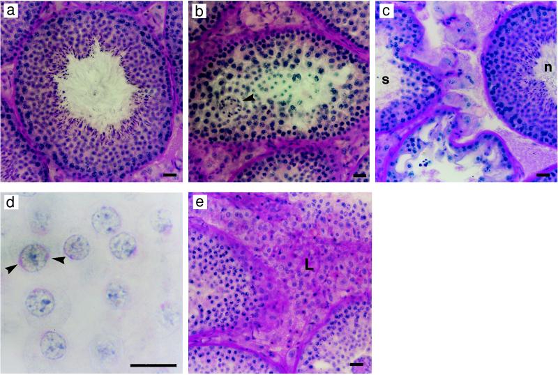
Testicular morphology. (a) All four 1-year-old w/t animals had testes with normal morphology. (b) Major disruptions to spermatogenesis were evident in the testes of 1-year-old ArKO mice, the site of disruption appearing to be early spermiogenesis with symplasts (arrowhead) and degenerating early spermatids visible. (c) Most animals also had some tubules with normal morphology (n), adjacent to tubules with spermiogenic arrest (s). (d) In tubules in which spermiogenic disruption was evident, there also appeared to be impaired acrosomal development. An example is shown in which spermatids with abnormal acrosome development are seen in a stage IV–V tubule. Multiple acrosomal vesicles were noted (arrowheads), and in some cases acrosomes failed to uniformly spread over the spermatid nuclei. (e) All animals showed evidence of what appeared to be Leydig cell hyperplasia/hypertrophy (L). (Scale bars: a–c and e, 20 μm; d, 10 μm.)

