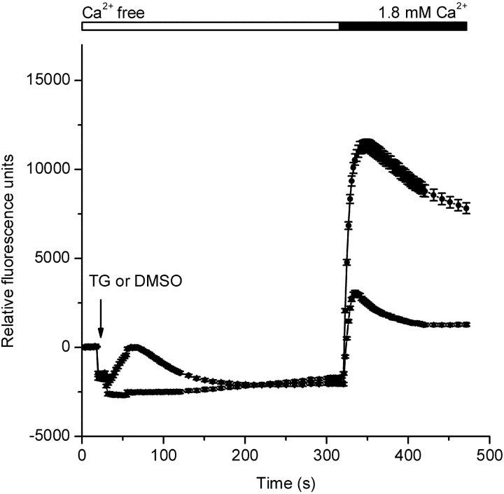
Figure 1.
Thapsigargin-dependent Ca2+ entry in S2 cells. Fluo-4 fluorescence changes were monitored using a FLIPR384. After 20 s of recording, the 384-well pipette-tip head was lowered into the solution creating an offset artifact in the recording. This offset artifact is unrelated to a cellular response and is dependent on the fluid volume in each well at the start of the experiment and the extent of tip penetration into the solution. 10 s after lowering the pipette-tip head, either thapsigargin (TG, 1 μM final, circles) or DMSO (triangles) was injected (arrow). CaCl2 was then added to achieve a final concentration of 1.8 mM. Traces were zeroed at time 0, and each data point represents the mean (±SEM) of 192 replicates.

