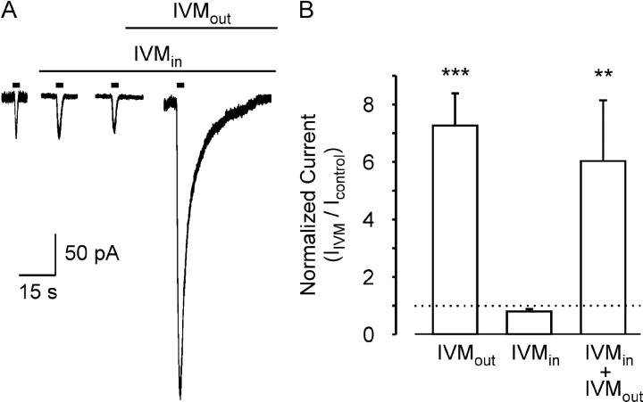
Figure 2.
IVM does not modulate hP2X4 receptor channels from within the cell. (A) Whole-cell current traces in response to ATP (3 μM) applied for 3 s every 3 min (short bars) before and after the addition of IVM (3 μM) to the extracellular solution, as indicated above the current traces. The first application of ATP was 20 s after establishing the whole-cell configuration. The standard pipette solution also contained IVM (3 μM). Holding potential of −50 mV. (B) Average (±SEM) amplitude of the whole-cell current activated by extracellular ATP (3 μM) 6 min after the addition of 3 μM IVM to the extracellular solution (IVMout), 6 min after exposure to intracellular 3 μM IVM (IVMin), or 5 min after the addition of 3 μM extracellular IVM to the cells exposed to intracellular IVM for 6 min (IVMin + IVMout). The amplitudes were normalized to the control response (dashed line). The bars represent between 5–14 cells. The statistical significance between IVMout and IVMin was determinate with the unpaired Student's t test, where *** represent P < 0.001, and between IVMin + IVMout and IVMin with paired Student's t test, where ** represent P < 0.01.

