
Figure 8.
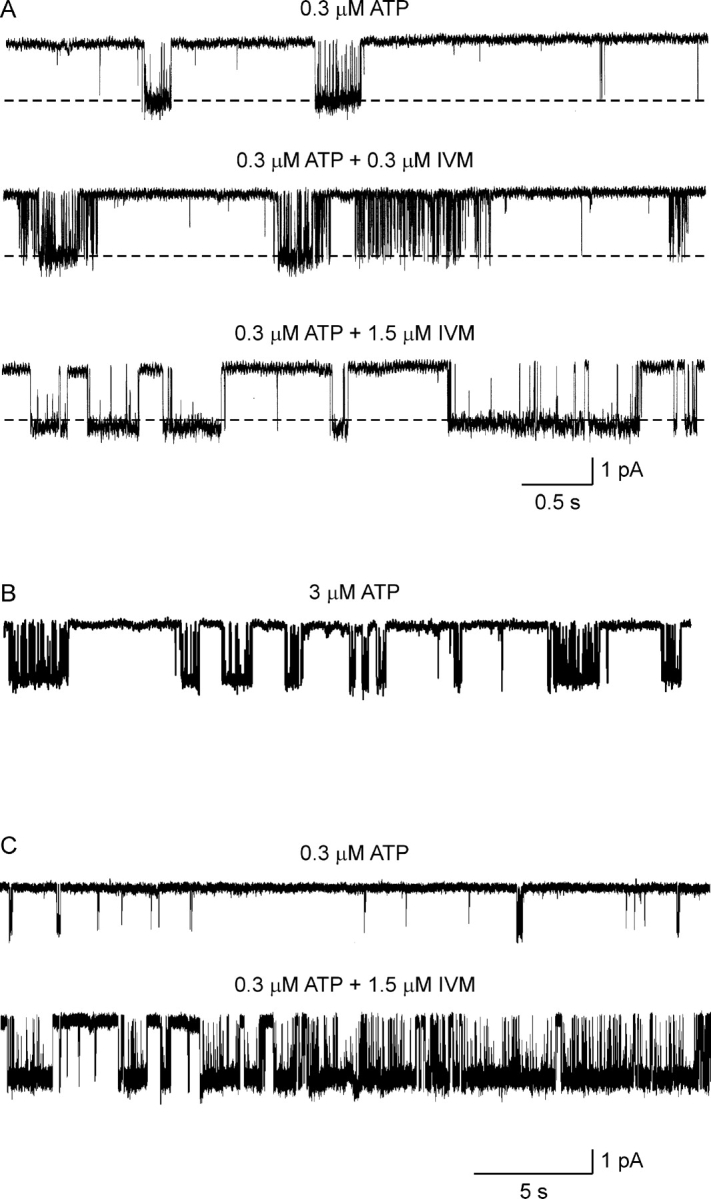
The effects of IVM on unitary hP2X4 receptor channel currents in on-cell patches. (A) Representative current records from three different on-cell patches. The pipette solution contained extracellular solution supplemented with 0.3 μM ATP (top trace), 0.3 μM ATP plus 0.3 μM IVM (middle trace), or 0.3 μM ATP plus 1.5 μM IVM (bottom trace). When IVM was included in the pipette solution, the cell was incubated in the same concentration of IVM for at least 25 min before establishing the cell-attached configuration. The dashed lines present the average current in the open state under control conditions. Sampled at 50 kHz and filtered at 1 kHz for display. (B) Representative single-channel current record with 3 μM ATP in the pipette solution. Same calibration as in A. Note the brief openings within the burst in comparison to the long openings induced by 1.5 μM IVM. (C) Representative current records 30 s in duration, from two different on-cell patches. The pipette solution contained extracellular solution supplemented with 0.3 μM ATP (top trace) or 0.3 μM ATP plus 1.5 μM IVM (bottom trace). Same experimental protocol as in A.