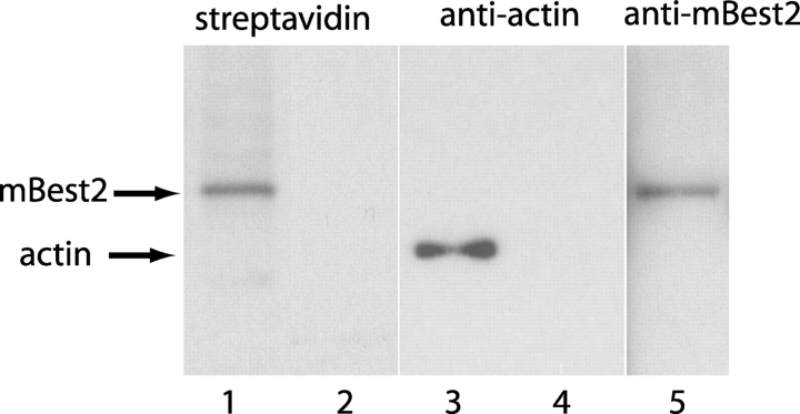
Figure 5.
Localization of mBest2 in the plasma membrane of HEK-293 cells. Lanes 1 and 2: streptavidin labeling of immunoprecipitated proteins. Nontransfected (lane 2) or mBest2-transfected (lane 1) HEK-293 cells were labeled with membrane-impermeant NHS-LC-biotin and lysed. mBest2 was immunoprecipitated with A7116 antibody, run on SDS-PAGE, and transferred to nitrocellulose. The nitrocellulose was then probed with HRP-conjugated streptavidin. A 64kD band was observed in transfected, but not nontransfected cells. Lanes 3–5: Western blots. Lane 3: total extract from mBest2-transfected cells was probed with antibody to α-actin. Lanes 4 and 5: the extract from mBest2-transfected cells was incubated with streptavidin-beads to collect biotinylated proteins. Biotinylated proteins were then probed with antibody to α-actin (Lane 4) or B4947 mBest2-specific antibody (Lane 5).

