
Figure 12.
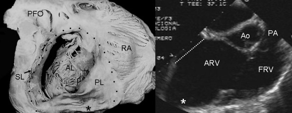
Internal view of right chambers of a specimen heart with Ebstein's anomaly shows significant dilatation at the atrioventricular junction and an aneurysmal fibrous sac on the right ventricular posterior wall (asterisk). The echocardiographic image shows the same finding (asterisk). Abbreviations as before.