
Figure 4.
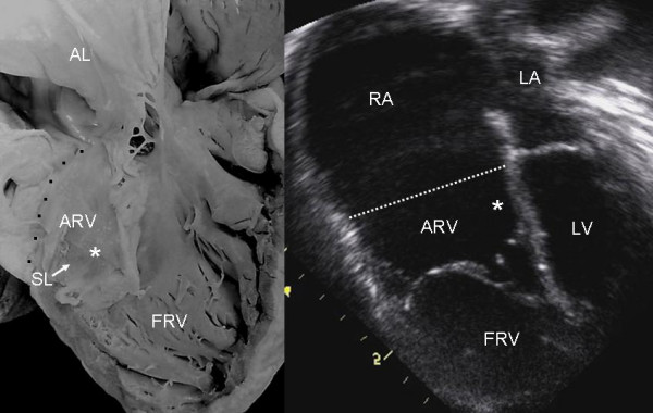
Internal view of the right ventricle shows grade II tethering of the tricuspid septal leaflet (asterisk). The 4 chamber echocardiographic image shows discontinuous leaflet tethering similar to the anatomic specimen. Abbreviations as before.