
Figure 7.
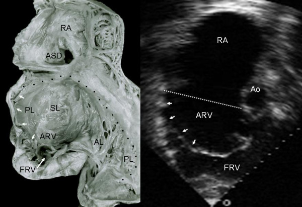
Internal view of the right chambers shows extreme tethering of the tricuspid septal and posterior leaflets to the ventricular wall (short arrows) and severe reduction of the functional portion of the right ventricle (long arrow). Note the atrial septal defect. The echocardiographic image shows the hammock-like aspect of the tethered posterior and septal leaflets that separate the atrialized and functional portions of the right ventricle. The atrialized portion of the right ventricle predominates. Ao: Aorta. Other abbreviations as before.