Figure 5.
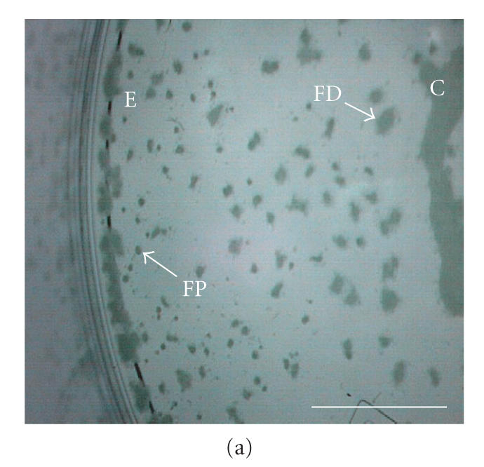
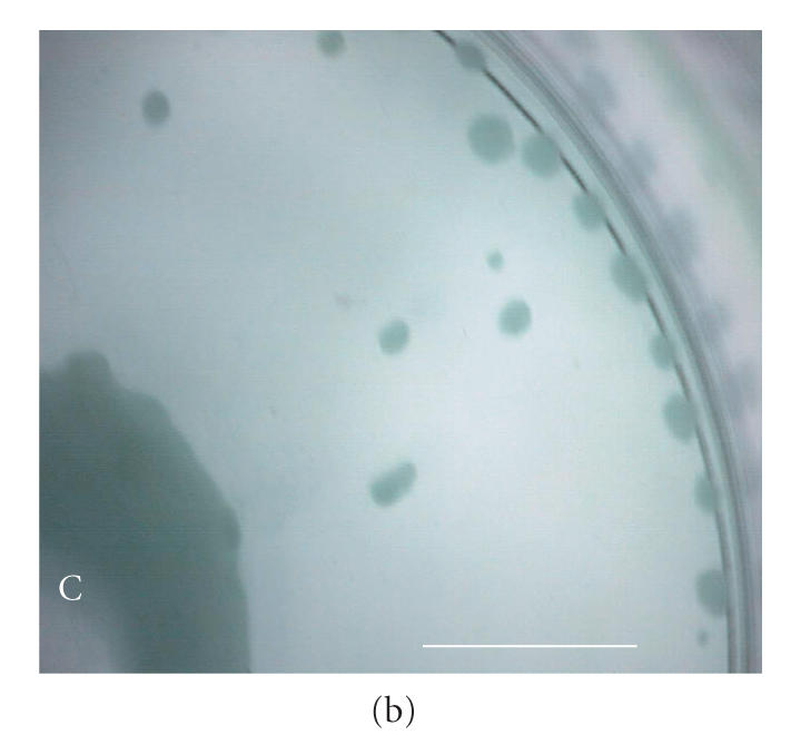
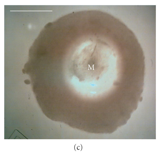
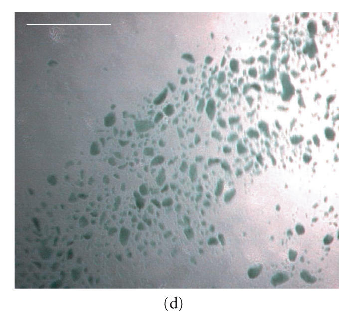
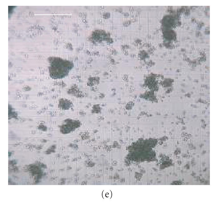
Micrographs of bioreactor cultures. (a) Base of a sector of an RDB bioreactor vessel following culture of EL-4 cells (without impeller agitation). EL-4 cells are dispersed across the surface as irregularly shaped plaques, with plaques merging into more confluent, continuous layers centrally (C) and along the circumferential edge (E), near the vessel side-wall. Intervening plaques located closer to the sidewall appear more focal punctate (FP) compared to more centrally located flattened discoid plaques (FD). (b) Base of a bioreactor vessel following culture of EL-4 cells with impeller agitation. Adherent plaques are larger discoid plaques, predominantly located centrally (C) and circumferentially (E), with a larger confluent tumor-like plaque with similarly smooth and flattened discoid appearance. (c) Magnified image of the central aggregate or “tumor” appearing in (b) showing a discoid, opaque colony with smooth edges. The centre of the plaque appears transparent due to a central dimple (manufacturing artefact) located on the plastic surface, a manufacturing artefact of the vessel over which a type of attenuated monolayer is visible (M). (d) Image of EL-4 aggregates of variable size, as viewed within a static tissue culture flasks. (e) Magnified image of EL-4 spheroid aggregates and disaggregated single cells contained on a hemocytometer slide, showing that spheroids are comprised of a homogeneous colony of densely packed, adherent cells. Scale bar = 10 mm (a)–(d) and 0.25 mm (e).
