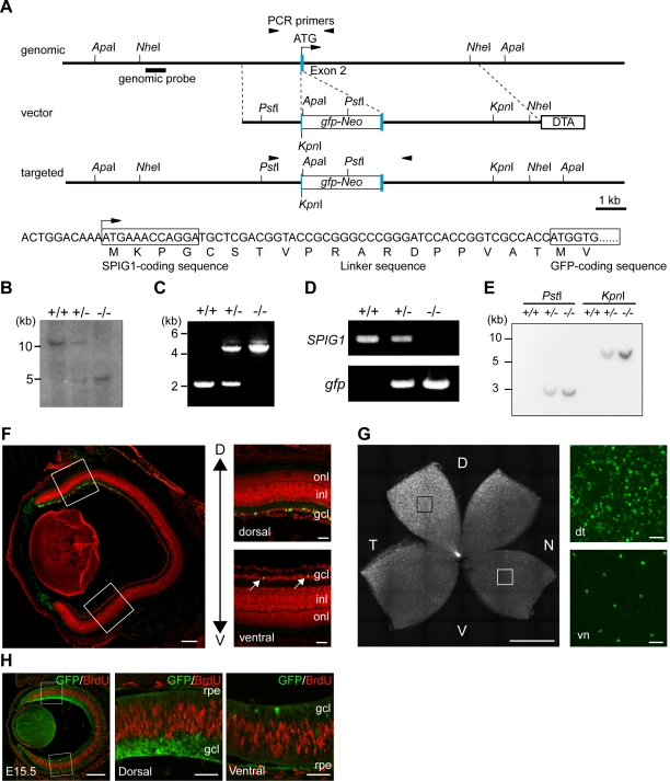
Figure 2. Generation of knock-in mice for the SPIG1 gene and expression of GFP in the retina.
A, Schematic representation of the structure of the endogenous allele (genomic), targeting vector (vector), and targeted allele (targeted). The genomic sequence of the head of exon 2 and the corresponding amino acid sequence in the targeted allele are shown below. The first coding exon of SPIG1 is indicated by blue boxes. For construction of the targeting vector, a gfp-Neo cassette was inserted in frame in the signal sequence of SPIG1 after the first four amino acids, Met-Lys-Pro-Gly, to yield fusion to the N-terminus of GFP through the linker sequence. The DT-A cassette was placed at the 3′ terminus of the homologous region for negative selection. The region used as a probe for Southern blotting is indicated by a bold bar. The position of primer sequences used for PCR analysis is also shown by arrowheads. B, Southern blot analysis of ApaI-digested genomic DNA from wild-type (+/+), heterozygous (+/−), and homozygous (−/−) mice. C, Genomic PCR analysis of wild-type (+/+), heterozygous (+/−), and homozygous (−/−) mice. D, RT-PCR analysis of total RNA from the brain of wild-type (+/+), heterozygous (+/−), and homozygous (−/−) mice. E, Southern blot analysis of PstI- or KpnI-digested genomic DNA from wild-type (+/+), heterozygous (+/−), and homozygous (−/−) mice with a probe for the coding region of gfp. F, Expression of GFP in the retina of SPIG1gfp/+ mice in a cross section at P5. Enlargements of the boxed regions are shown on the right. In the ventral area, GFP is expressed only in a small subset of cells in the GCL (arrows). In a cross section, dorsal (D) is upwards, and ventral (V) is downwards. inl, inner nuclear layer; onl, outer nuclear layer. G, Expression of GFP in the retina of SPIG1gfp/+ mice in a flat-mount at P5. The retina is apparently subdivided into two domains with SPIG1 expression, the dorsotemporal and pan-ventronasal domains. Enlargements of the boxed regions are shown on the right. d, dorsal; n, nasal; t, temporal; v, ventral. In the pan-ventronasal domain, SPIG1-expressing cells form a regular mosaic. H, Double staining of GFP and BrdU in a cross section at E15.5. Enlargements of the boxed regions are shown on the right. GFP is not expressed in the proliferating cells at S phase. rpe, retinal pigment epithelium. Scale bars: 200 µm (left panels in F/H); 50 µm (enlargements in F/G, and H); 1 mm (left panel in G).

