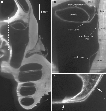
Fig. 2.
(a) 2D OPFOS-image of the region around Bast’s valve and the utricular duct, obtained with another OPFOS setup [7] than for (c) and following. The dashed box is shown in a larger magnification in (b), (b) Dashed box in (a) shown in a larger magnification, (c) 2D OPFOS-image of Bast’s valve. The arrows point toward very narrow passages

