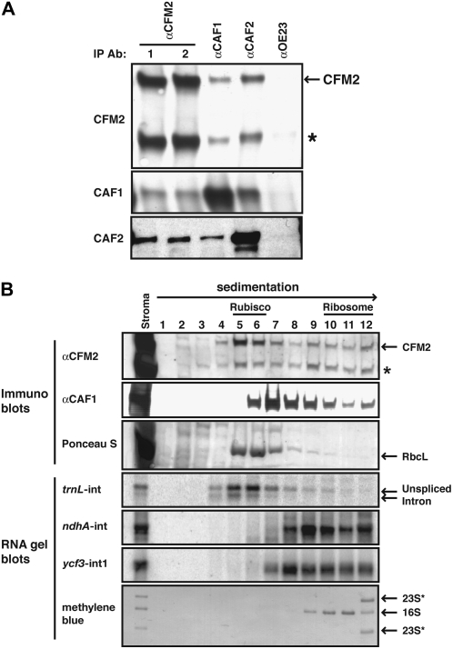
Figure 4.
CFM2 Is Found in Large RNPs That Include CAF1 and/or CAF2.
(A) Immunoblots showing coimmunoprecipitation of CFM2 with CAF1 and CAF2. Immunoprecipitations were performed with chloroplast stroma, using the antibody named at the top (IP Ab); antibodies from two different rabbits immunized with the CFM2 antigen were used in replicate assays (lanes 1 and 2). The presence of specific proteins in the immunoprecipitation pellets was determined by probing replicate blots with the antibodies listed to the left. All antibodies were affinity purified against their cognate antigen prior to use. An anti-OE23 immunoprecipitation served as a negative control. The asterisk marks the ∼80-kD protein detected with the CFM2 antisera.
(B) Cosedimentation of CFM2 with CAF1, CAF2, and intron RNAs. Stromal extract was sedimented in sucrose gradients under conditions in which particles greater than ∼60S pellet. An equal proportion of each fraction was analyzed by probing immunoblots with the antibody indicated to the left. The Ponceau S stained blot illustrates the position of Rubisco in the gradient; RbcL is the large subunit of Rubisco. Bottom panels show RNA gel blots of RNA purified from the same gradient fractions. The probes were intron specific and are indicated to the left. The methylene blue–stained blot illustrates the position of ribosomal subunits in the gradient: 16S rRNA marks 30S ribosomal subunits, and 23S* rRNAs mark 50S ribosomal subunits.

