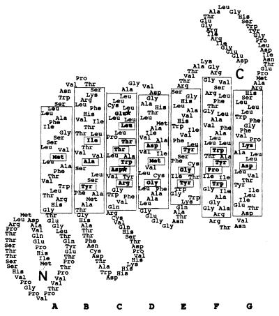
Figure 2.
Predicted topology of the NOP-1 protein. The seven transmembrane α-helices are designated A through G, and helix boundaries are based on those of BR (18). Arg-128 in helix C has been included in the pocket in accordance with the crystal structure of BR (30). Retinal-binding pocket residues conserved among the archaeal transport and sensory rhodopsins (31, 32) are boxed. ∗ mark positions homologous to Asp-85 and Asp-96 in BR. There are two in-frame methionines upstream of helix A; it is not known whether one or both are used for translation.

