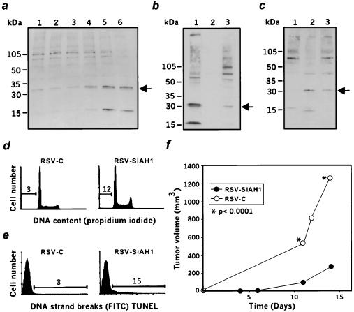
Figure 2.
Characterization of SIHA1. (a) Expression of the murine siah-1b in the course of p53-induced apoptosis, as measured by Western blot analysis with anti-SIAH-1. LTR-6 cells at 37°C (lane 1) and after 4 hr, 7 hr, 9 hr, 16 hr, or 24 hr of incubation at 32°C (lanes 2–6). Arrow indicates the 32-kDa siah-1b protein. (b) Subcellular localization of murine siah-1b in LTR-6 cells determined by Western blot analysis with anti-SIAH-1 antibodies. Lanes 1, 2, 3 represent the nuclear, membrane, and cytoplasmic fractions respectively. (c) Expression of SIAH-1 in human U937 cells. Lane 1, U937 cells transfected with the control vector. Lane 2, US cells derived from U937 cells but displaying a suppressed malignant phenotype. Lane 3, U937 cells stably transfected with SIAH-1. (d and e) Fluorescence-activated cell sorting analysis of DNA content (d) or TUNEL staining (e) in U937 cells transfected with the vector (RSV-C) or transfected with SIAH-1 (RSV-SIAH-1). (f) Tumorigenicity assay in scid/scid mice after injection of U937 cells transfected with either the control vector (○, RSV C) or with SIAH-1 (●, RSV-SIAH-1).

