Figure 4.
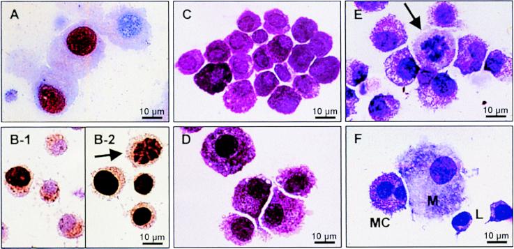
Ki-67 expression, BrdUrd incorporation, and morphology of human MC. (A) Immunostaining of MC (purity 98%) cultured with IL-4 and SCF for 14 days. The nucleus of MC that express the proliferation marker Ki-67 is colored red. (B) BrdUrd incorporation by MC (dark brown staining) cultured for 4 days (B-1) or 14 days (B-2, arrow indicates a mitotic figure) in the presence of IL-4 and SCF. (C) Isolated human intestinal MC before culture (MC purity 81%). (D) Same cells as in C after 14 days of culture with SCF and IL-4. MC were enlarged and had more abundant cytoplasmatic projections. MC purity increased to 99%. No morphological differences were observed between MC cultured with SCF alone (not shown) or MC cultured with SCF and IL-4. (E) Occasionally, mitotic figures (arrow) could be detected in cultures supplemented with SCF alone or SCF and IL-4. (F) Double-lobed MC developed in vitro from PBMC cultured as described in the text. The MC are neighbored by a macrophage and two lymphocytes. Cells were stained with May-Grünwald/Giemsa (C–F).

