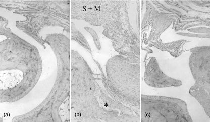
Fig. 2.
Histology from the right hindleg joint of C3H mice. (a) Normal control mouse. Ordinary synovia and preserved cartilage–bone interface. (b) Sixteen days after inoculation with Borrelia burgdorferi. Hyperplasia of synovia (S) with massive infiltration of mainly mononuclear inflammatory cells (M). Cartilage destroyed with superficial erosion of the bone structure (*). (c) Sixteen days after inoculation of B. burgdorferi and 27 days after onset of mercury treatment. Minimal synovial hyperplasia with scant inflammatory cells and preserved cartilage–bone interface.

