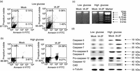
Fig. 2.
IA-2 transfected mouse insulinoma MIN-6 cells undergo apoptosis in high glucose media. (a, b) Flow cytometry detection of apoptotic cells as determined by uptake of propidium iodide and by staining with anti-annexin V-fluorescein isothiocyanate (FITC) in (a) low and (b) high glucose media in mock and IA-2F transfected cells. (c) DNA fragmentations of cells in low and high glucose media. (d) Caspase-3, 6, 7, 9, 12 and caspase activated DNase (CAD) activity detected by Western blot in low and high glucose media.

