Figure 2.
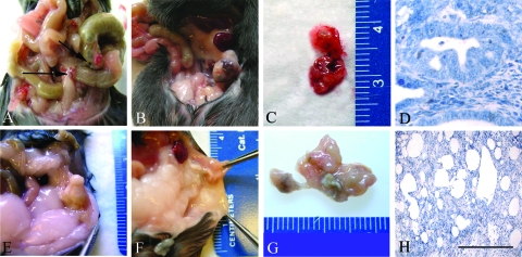
Endometriosis-like lesions in Leprdbm (A–D) and Leprdb hosts (E–H). After uterine tissue transfer, each host type received uterine tissue from the female siblings of the opposite genotype of the same litter. Seven days after transfer, Leprdbm animals (received Leprdb tissue) displayed multiple vascular lesions (A), compared with the purulent nonvascular sites in Leprdb hosts (received Leprdbm tissue) (E). In long-term studies (14 d after transfer), lesions of the Leprdbm hosts appeared significantly more vascularized grossly and invasive at multiple sites (B and C) in contrast to Leprdb hosts, which did not display significant vascularization or mottling, and demonstrated marked purulence and necrosis (F and G). Microscopic comparison (hematoxylin and eosin) revealed glandular architecture and organization in Leprdbm hosts (D) similar to that seen in the WT syngeneic transfer model. Leprdb showed diffuse necrosis and glandular and stromal disruption (H). Microscopic images (hematoxylin and eosin), ×60. Scale bars, 100 μm.

