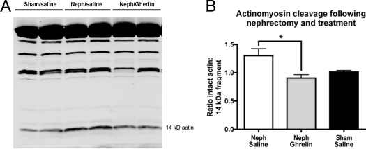
Figure 8.
Muscle actinomycin degradation. A, Representative Western blot of calf muscle lysate from Neph animals receiving saline or ghrelin and sham-operated rats receiving saline. Western blot was performed using an antibody to the carboxy terminus of actin, showing the 42-kDa intact actin band (top band) and the cleaved 14-kDa fragment (labeled). B, Ratio of intact actin to 14-kDa fragment for Neph/saline, Neph/ghrelin, or sham/saline animals. Significance is indicated: *, P < 0.05.

