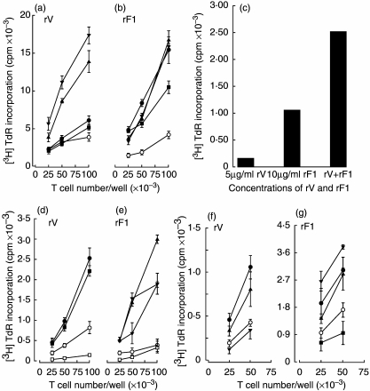
Fig. 4.
Primary T cell proliferative responses in vitro to rV and rF1 antigens. Different numbers of lymph node T cells from naive mice were cultured with 1000 autologous dendritic cells (DC), which were either unpulsed or pulsed with antigens. Proliferation was measured on day 3 of culture. (a, b) Three-day bone marrow-derived DC (BMDC) were either unpulsed (O) or pulsed with rV (a) or rF1 (b) at 0.1 (▪), 0.5 (•), 5 (▴), 10 (▾) for 18 h. (c) Naive donor T cells (105) were cultured with splenic DC pulsed with 5 µ g/ml rV, 10 µg/ml rF1 or both antigens at these same concentrations for 18 h. (d–e) Seven-day BMDC. Responder cells only (□). DC were either unpulsed (O) or pulsed with rV (c) at 5 (▪) and 10 µg/ml (•) or rF1 (d) at 1 (▴), 5 (▾) and 10 µg/ml (★) for 2 h. (f–g) Splenic DC were either unpulsed (O) or pulsed with rV (e) at 2.5 (▾), 4.5 (▴) and 10 µg/ml (•) for 2 h; or rF1 (f) at 5 (▪), 10 (•), 20 (▴) and 25 µg/ml (▾) for 4 h. All figures are representative of data gained in at least three experiments.

