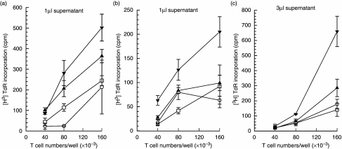
Fig. 5.
Primary T cell proliferative responses to supernatants from rV- and rF1-pulsed DC. (a) Cell free supernatants were collected at 24 h of culture from 3d bone marrow-derived DC (BMDC) which had been pulsed with rV and rF1 for 4 h, and then washed. The supernatants (1 µl) were used to stimulate syngeneic lymph node T cells. Lymph node cells alone (□); supernatants from unpulsed DC (O); supernatants from rV- and rF1-pulsed DC (▴) and (▾), respectively. (b, c) Cell free supernatant (b: 1 µl, c: 3 µl) from splenic DC pulsed for 2 h with rV and rF1 and used to stimulate autologous lymph node T cells. Legends for data sets are represented as in (a).

