
Fig. 6.
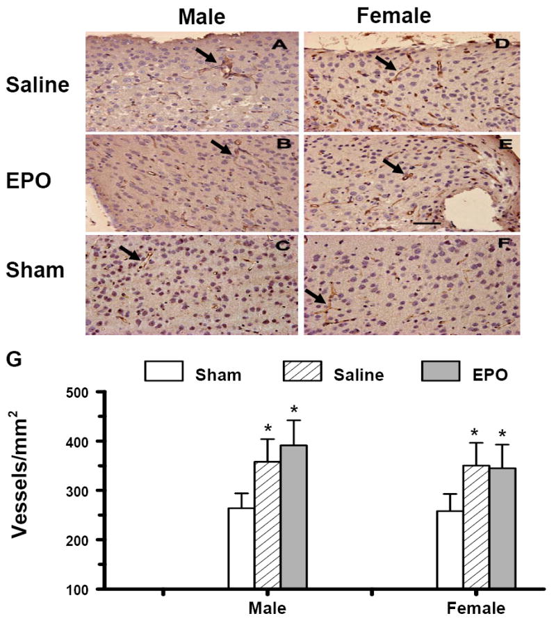
Effect of rhEPO on vWF-staining vascular structure in the ipsilateral cerebral cortex of sham or injured mice in both genders at 35 days after TBI. TBI (A, D) alone significantly increased vascular density (stained brown, arrow as an example) in the cortex. rhEPO (B, E) did not affect angiogenesis after TBI. The density of vWF-stained vasculature is shown in (G). Data represent mean ± SD. *P < 0.05 vs. Sham. N (mice/group) = 13 (male-saline), 12 (male-EPO), 8 (male-sham), 8 (female-saline), 8 (female-EPO), 7 (female-sham). Scale bar = 50μm.