
Fig. 7.
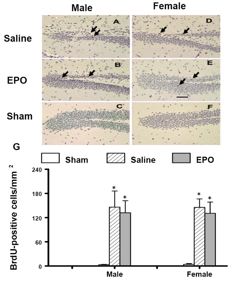
Effect of rhEPO on BrdU-positive cells in the ipsilateral DG at 35 days in both genders after TBI. TBI (A, D) alone significantly increased the number of BrdU-positive cells (brown stained, arrows) in the ipsilateral DG. However, treatment with rhEPO (B, E) did not affect cell proliferation as compared with the saline group (A, D). The number of BrdU-positive cells is shown in (G). Data represent mean ± SD. *P < 0.05 vs. Sham. N (mice/group) = 13 (male-saline), 12 (male-EPO), 8 (male-sham), 8 (female-saline), 8 (female-EPO), 7 (female-sham). Scale bar = 50μm.