
Fig. 5.
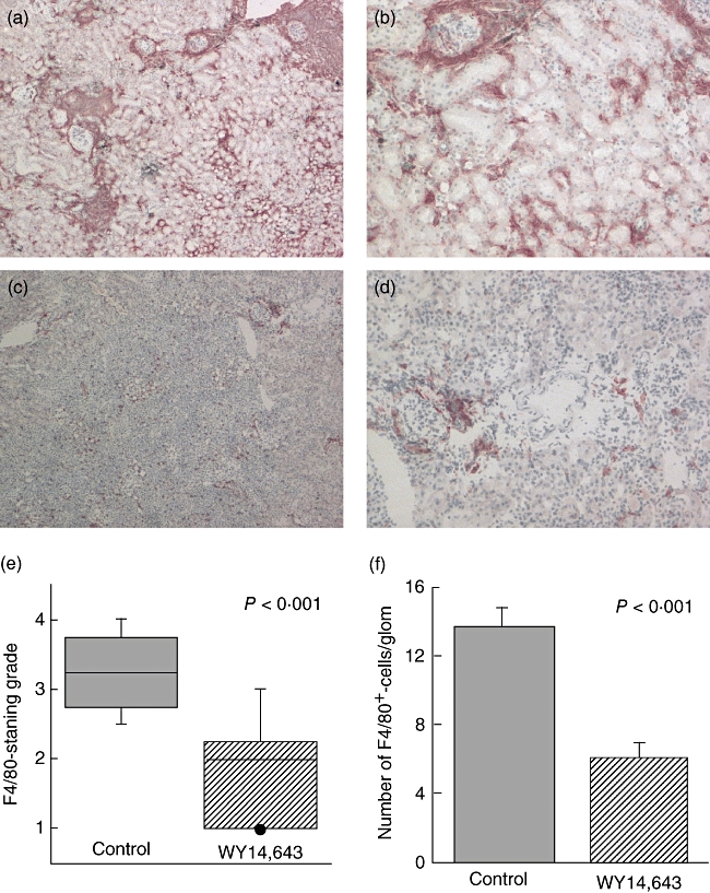
(a) A PPARα ligand decreases renal macrophage infiltration. Frozen sections were stained with the macrophage marker F4/80 and immunohistochemistry was performed. (a) and (b) Representative sections of F4/80-staining of control and (c) and (d) sections of WY14,643-treated mice (a and c, 40×; b and d, 100×). (e) The histological grade was significantly lower in the PPARα-ligand-treated animals (P < 0·001, Mann–Whitney U-test; N = 27). (f) The number of F4/80+-staining cells per glomerulus was also determined and there was a significant decline in the number of intraglomerular macrophages in the WY14,643-treated mice. P < 0·001, Student's t-test; N = 27.