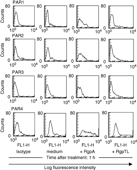
Fig. 5.

Flow cytometric analysis of the effect of RgpA on the surface expression of the protease-activated receptor (PAR) family in T cells. T cells were stimulated with medium alone or medium containing 70 nM RgpA; or N-α-tosyl-L-lysyl chloromethyl ketone (TLCK)-inhibited RgpA (RgpTL) for 1 h at 37°C. After incubation, cells were stained with isotype-matched control or anti-PAR-1, -PAR-2, -PAR-3 and -PAR-4 followed by flow cytometry.
