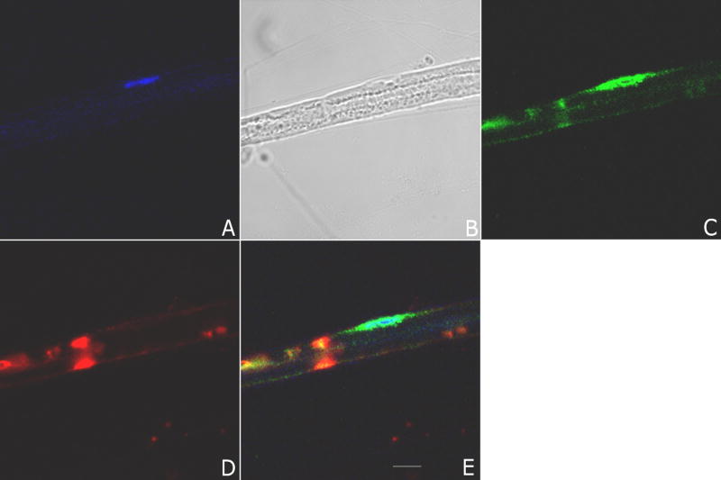
Figure 1. Confocal microscopy of myelinated teased fiber preparations from rat sciatic nerve.
Panel A shows TOPRO-3, a nuclear stain (blue), and the presence of one Schwann cell nucleus; Panel B shows a phase contrast image of the teased fiber; Panel C shows immunofluorescence with polyclonal antibody to a particularly interesting new cysteine histidine protein (PINCH) (Alexa 488, green); Panel D shows immunofluorescence using anti-myelin associated glycoprotein (MAG) (Alexa 564, red), a marker of Schwann cell cytoplasm where myelin is not compact; Panel E shows colocalization of MAG, PINCH and TOPRO-3. Note the peri-nuclear staining of PINCH and localization in compartments of non-compact myelin. Images represent at least 10 separate fibers analyzed. Scale bar 10 μm.

