Figure 1.—
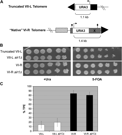
Telomere structure and TPE analysis at the truncated VII-L and VI-R telomeres. (A) The VI-R subtelomere contains a 380-bp “core X” element (shaded), containing an ARS consensus sequence (ACS; circle) and Abf1p binding site (diamond). The URA3 TPE reporter was introduced at the VI-R telomere X-ACS in a manner analogous to the creation of native TPE reporters described in Pryde and Louis (1999). PCR primers whose 5′ ends corresponded to VI-R X-element sequence surrounding the X-ACS were used to amplify URA3 from ADH4UCAIV, the same plasmid used to create the truncated VII-L telomere reporter (Gottschling et al. 1990). Unlike the system that truncated the VII-L telomere at ADH4 (solid), the “native” VI-R TPE reporter preserves the subtelomeric structure. The closest upstream gene is YFR057w (striped box). Both strains also contain upstream lac operator arrays for visualization studies (Mondoux et al. 2007). All strains used in this study were constructed in the YPH background (ura3-52 lys2-801 ade2-101 trp1-Δ63 his3-Δ200 leu2-Δ1; Sikorski and Hieter 1989) and grown at 30° in yeast complete (YC) synthetic medium and plates (Zakian and Scott 1982). All TPE reporter strains were verified for correct integration by Southern blotting and pulsed-field gel electrophoresis. SIR1 was deleted in each strain background using a PCR-mediated knockout that eliminated the complete open reading frame, replacing it with a hygromycin resistance cassette (Goldstein and McCusker 1999). (B) TPE assays. Tenfold serial dilutions of the VI-R, truncated VII-L, and sir1Δ versions of these strains were plated onto +Ura or 5-FOA plates to assay silencing and photographed after 3 days' growth. TPE is higher at VI-R compared to truncated VII-L and is not dependent on Sir1p at either telomere. (C) Quantitation of TPE. +Ura-grown cells were plated onto +Ura and 5-FOA plates and colonies counted after 3 days of growth. The percentage of total cells (+Ura) that grew on 5-FOA plates is represented as % TPE. Error bars represent standard deviations. TPE at VI-R is significantly higher than TPE at VII-L by Student's t-test (P < 7 × 10⁻⁵). There was no significant difference in TPE at either the VI-R or VII-L telomere in the absence of Sir1p.

