
FIG. 3. Unloading of fibrous-bony insertion sites by tail suspension (A–C) or surgical transection (D–K).
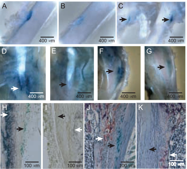
(A and B) Sham control (A) and suspended (B) insertions of the adductor magnus into the mid-posterior femur in a 7-week-old male PTHrP-lacZ mouse suspended for 11 days, cleared images of X-gal-stained specimens (50X). (C) Origin of the vastus lateralis (arrows) from the third trochanter of a 7-week-old mouse suspended (image to left) or shamed (to right) for 7 days. (D and E) Control (D) and transected (E) MCL/SM insertions (arrows) in a 4-week-old PTHrP-lacZ mouse sacrificed 7 days following transection, 40X. (F and G) Control (F) and transected (G) semitendinosus insertions (arrows) in a 4-week-old mouse sacrificed 7 days following transection, 40X. (H and I) Control (H) and transected (I) MCL insertions in the left and right legs, respectively, of a 5-week-old PTHrP-lacZ mouse 7 days after surgery, X-gal and alkaline phosphatase stains. The closed arrow identifies β gal-positive cells and the open arrow alkaline-phosphatase activity. The images focus on the trailing edge of the insertion. The ratio of unloaded: control integrated greyscale values for the alkaline phosphatase-stained areas was 0.25 ± 0.03 (mean ± SE, p<0.001). (J and K) Control (J) and transected (K) MCL insertions contiguous to those shown in panels H and I, X-gal and TRAP stains. The closed and open arrows identify β gal- and TRAP-positive cells, respectively. The images focus on the leading edge of the MCL, flanked internally by the trabecular bone of the tibial metaphysis. The mean (± SE) numbers of TRAP-positive cells fell from 114 ± 23 in the control to 22 ± 8 cells/µm² in the transected sites (p<0.001).