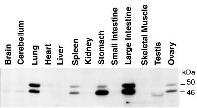
Figure 1.
Analysis of VASP expression in wild-type mice. Cell homogenates (30 μg protein per lane) were probed with an affinity-purified VASP polyclonal antibody. Equal protein loading was controlled by Ponceau staining. VASP appears as a doublet of 46 kDa and 50 kDa representing the serine-157 dephosphorylated and phosphorylated protein states, respectively. Longer exposure of this immunoblot also demonstrated VASP expression in brain, heart, kidney, and small intestine.

