
Fig. 2.
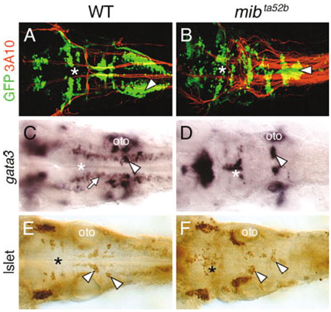
Branchiomotor neuron (BMN) development is severely disrupted in the mind bomb mutant hindbrain. All panels show dorsal views of the hindbrain with anterior to the left. A and B are composite confocal images and identify GFP-expressing motor neurons in the fluorescein channel and 3A10-labeled Mauthner (M) reticulospinal neurons and axons in the rhodamine channel. The asterisks in all panels indicate the location of the trigeminal motor neurons in rhombomeres 2 and 3. A,B: In a 36 hpf wild-type embryo (WT; A), the BMN clusters are found in their characteristic locations and numbers. The 3A10 antibody-labeled Mauthner cell axons decussate and descend contralaterally into the spinal cord. In a mib mutant (B), The BMNs clusters are disorganized and are variably fused. The supernumerary M cell axons cross the midline and descend contralaterally in a normal manner, whereas the nX motor neurons (arrowhead) in the same region exhibit extensive fusion across the midline. C,D: In a 36 hpf wild-type embryo (C), gata3 is expressed by putative interneurons (arrow) and the nVII (arrowhead) and nV motor neurons (asterisk). In a mib mutant (D), gata3-expressing motor neurons (arrowhead, asterisk) are disorganized and the putative interneurons are absent. E,F: In a 36 hpf mib mutant (F), islet antibody-labeled BMNs (asterisk, arrowheads) are disorganized and variably fused and are reduced in number compared with wild-type siblings (E). oto, otocyst.