
Figure 3.
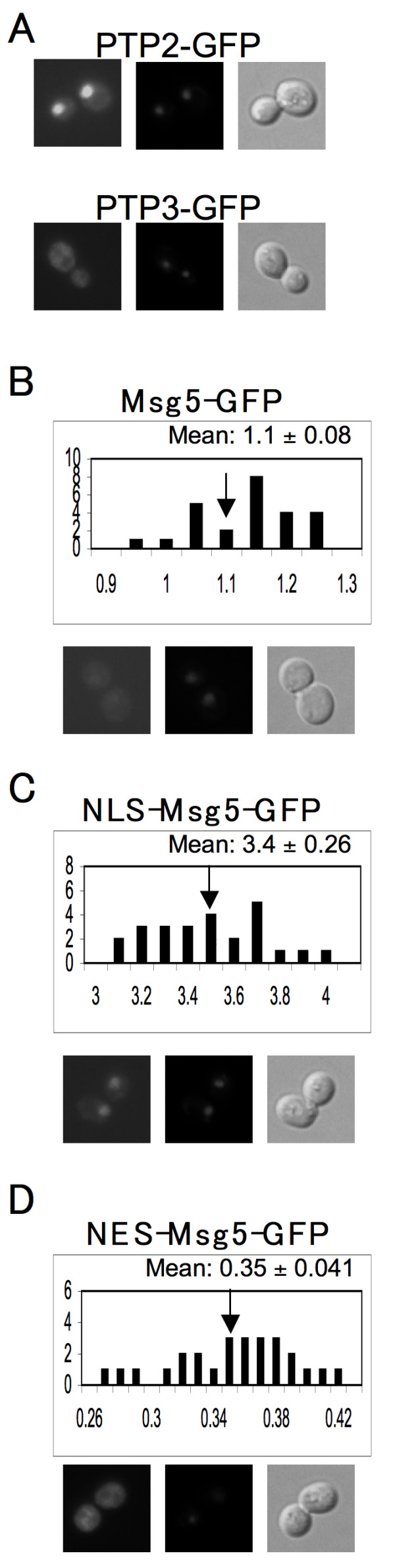
Localization of the wild type, NLS-tagged, and NES-tagged forms of Msg5. A set of 15Dau transformants carrying various centromeric reporter plasmids–PTP2-GFP, PTP3-GFP, MSG5-GFP, NLS-MSG5M45A-GFP, and NES-MSG5M45A-GFP–were grown to mid-log phase, stained with 20 μg/ml DAPI, and examined using fluorescent and differential interference contrast (DIC) microscopy. In each panel, representative images are displayed as follows: FITC (left), DAPI (middle), and DIC (right). RNCF values for Msg5-GFP, NLS-MSG5M45A-GFP, and NES-MSG5M45A-GFP were quantified in the same manner as for Fus3-GFP, and are represented as histograms. RNCF values are indicated as the mean ± s.d. (A)PTP2-GFP cells (top); PTP3-GFP cells(bottom); (B)MSG5-GFP cells; (C)NLS-MSG5M45A-GFP cells; (D)NES-MSG5M45A-GFP cells.