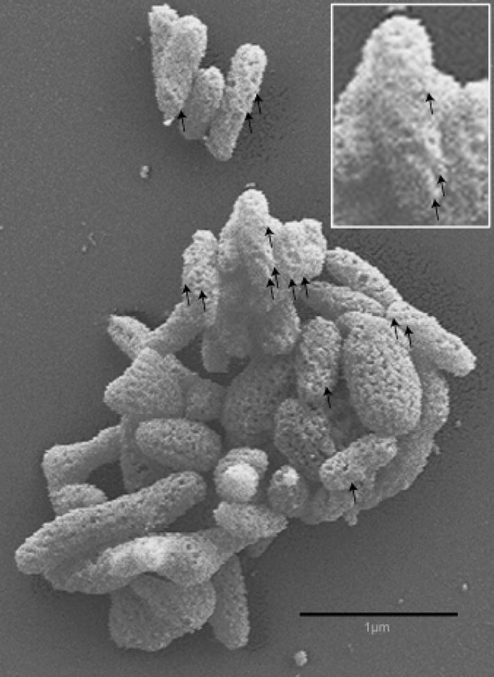
Fig. 2.

Scanning electron micrograph of Mycoplasma insons I17P1Tcells showing a twisted rod morphology (inset), absence of the coiled symmetry of spiroplasmas, and a highly textured surface. Raised ridges following a helical path along the surface are prominent on some cells (arrows).
