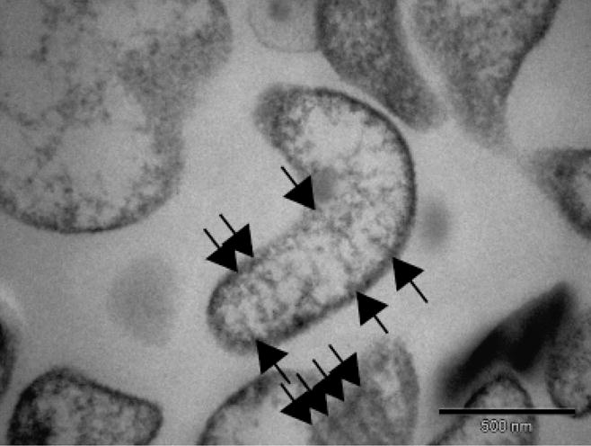
Fig. 3.

Transmission electron micrograph of a thin section of Mycoplasma insons I17P1T cells showing a single cytoplasmic membrane, absence of cell wall, and absence of a tip structure. Diagonal striations across the cut cell sections (arrows) were similar to Mycoplasma fastidiosum (Lemcke & Poland, 1980).
