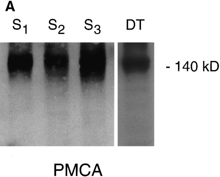
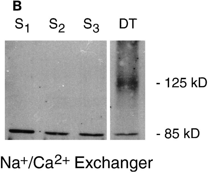
Figure 5.
Analysis of PMCA and Na+/Ca2+ exchanger protein expression in proximal tubule cells. Western analysis was performed on proximal tubule cell membrane preparations. Lane 1, S1 membranes; lane 2, S2; lane 3, S3; and lane 4, primary cultures of mouse distal tubule cells. The same preparations were probed with either: (A) an mAb against PMCA, which revealed a reacting protein of ∼140 kD; or (B) a polyclonal antibody raised against a fusion protein encoding the rabbit kidney exchanger. Bands of 125 and 85 kD, were seen in the positive control, distal tubule cells, whereas the proximal tubule cell membrane preparations revealed only an 85-kD band.


