Figure 1.
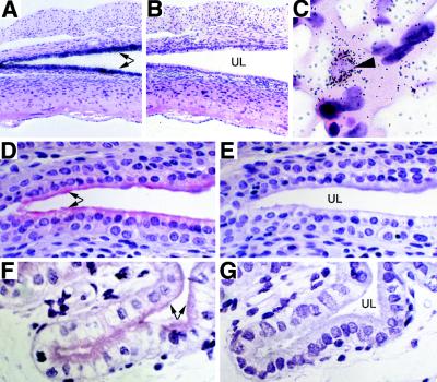
TF expression in the uterus and placenta. In situ hybridization experiments were performed to determine mouse TF mRNA expression in the uterus and placenta (E13.5) of a wild-type mouse (A– C). Uterine sections from a wild-type mouse were incubated with antisense (A) or sense (B) mouse TF riboprobes (×100). (C) The labyrinthine zone of a wild-type placenta incubated with a mouse TF antisense riboprobe (×1,000). Arrowhead indicates a mouse TF mRNA-positive trophoblast within the placental labyrinth. Immunohistochemical analysis was used to localize mouse and human TF in the uterus of wild-type and low-TF mice, respectively. Uterine sections from a wild-type mouse were incubated with a sheep anti-rabbit TF polyclonal antibody (D) or a control antibody (E) (×400). Uterine sections from a low-TF mouse were incubated with a sheep anti-rabbit TF polyclonal antibody (F) or a control antibody (G) (×400). Arrows indicate TF-positive uterine epithelium. UL, uterine lumen.

