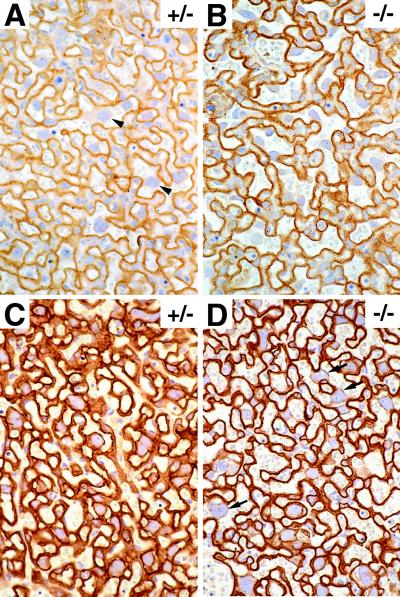
Figure 4.
Immunohistochemical analysis of the placental labyrinth in TF null and normal TF (+/−) placentas. Placental sections from normal TF (A and C) and TF null (B and D) embryos were incubated with an anticadherin antibody (A and B) or an anticytokeratin antibody (C and D). Arrowheads identify layer I trophoblasts with multiple cellular contacts to adjacent trabeculae in normal (+/−) placentas (A). Note the larger maternal lacunae and reduced cytokeratin staining in layer I trophoblasts of TF null placentas (arrows in D) compared with the placentas from normal (+/−) TF placentas. (×400.)

