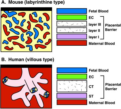
Figure 6.
Structure of the maternofetal barrier in mouse and human placentas. The mouse and human placentas have labyrinthine and villous types of interdigitation between maternal and fetal tissues, respectively. Maternal blood is red, fetal blood is blue, and fetal trophoblasts are black. Mice and humans form hemotrichorial placentas. In mice, the maternofetal barrier consists of four layers: (i) a fetal endothelial cell layer (EC), (ii) trophoblast layer III, (iii) trophoblast layer II, and (iv) trophoblast layer I. In humans, three layers are present: (i) a fetal endothelial cell layer (EC), (ii) a cytotrophoblast layer (CT), and (iii) a syncytialtrophoblast layer (ST).

