
Figure 5.
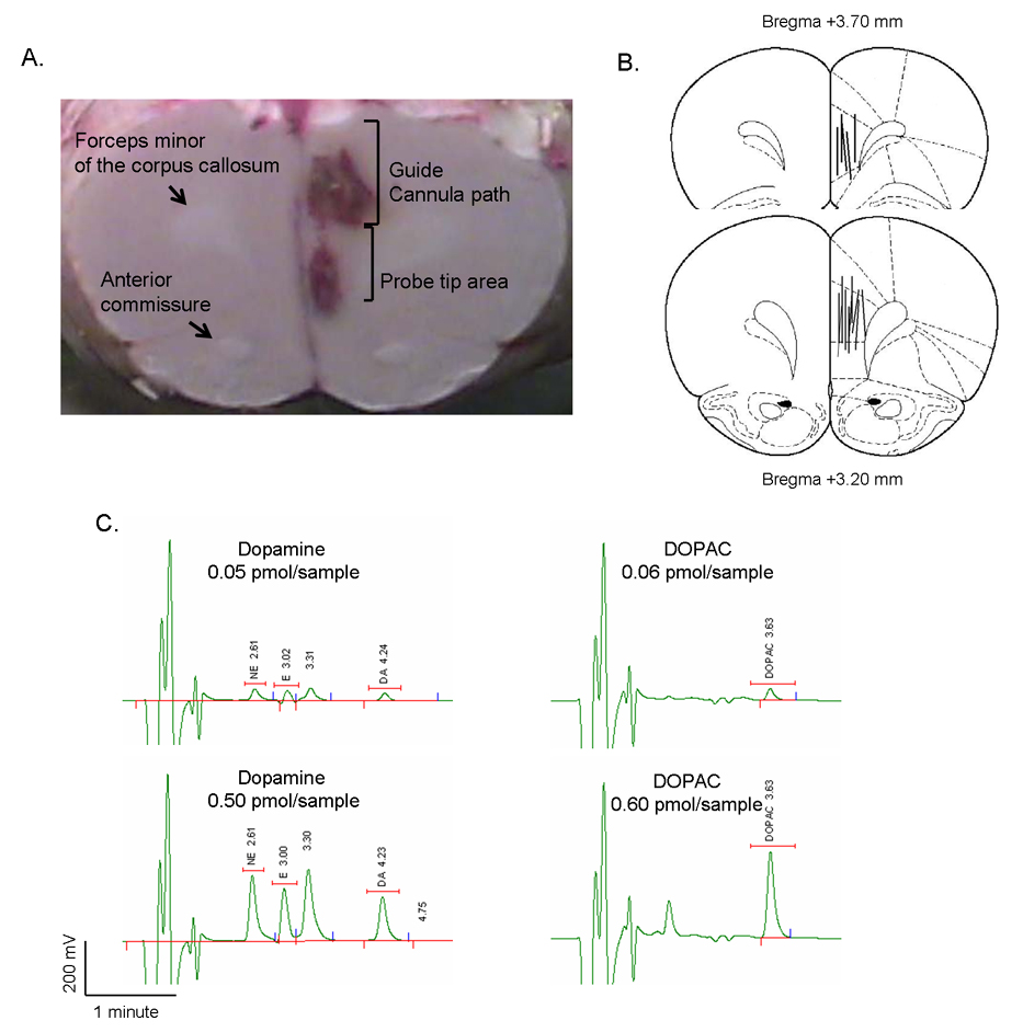
Microdialysis experiments. A: Histological identification of the guide cannula placement into the medial prefrontal cortex. Representative fresh rat brain sectioned after microdialysis experiments. Gross anatomical localization was used to assess correct cannula placement, using the forceps minor of the corpus callosum as a reference landmark. Guide cannula trace and probe tip area are discernable. B: Paxinos and Watson atlas maps indicating site of cannula placement across all animals. C: Chromatograms showing retention times for two concentrations of dopamine and DOPAC.